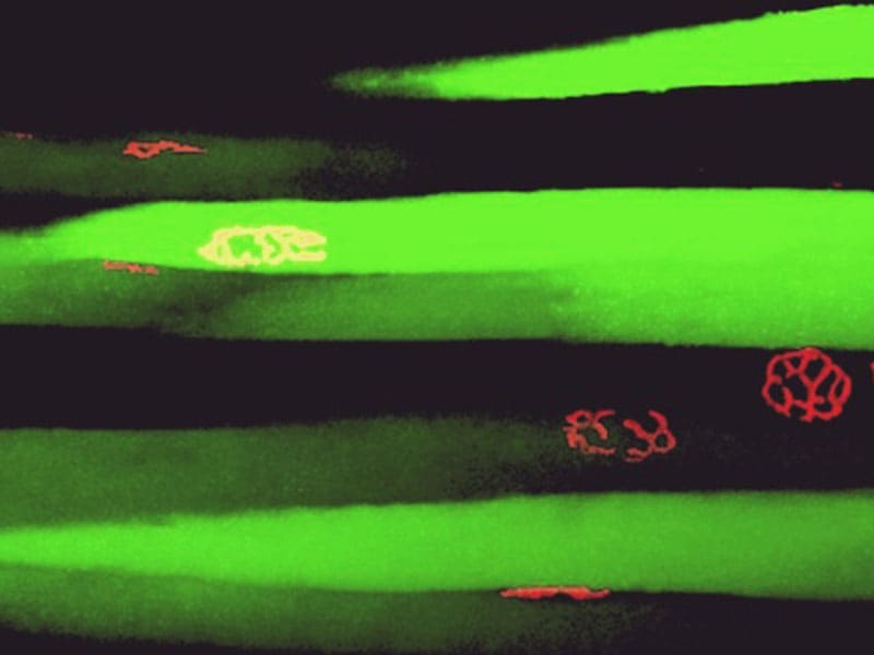
Skeletal Muscle Tissue from Tibialis Anterior of a Young Adult Mouse
Neuromuscular junction structure stained with α-bungatotoxin Alexa Fluor 555 dye (red). Gene
activity for shRNA of CAV1 gene stained with GFP green florescent protein (green).
Courtesy of José G. Grajales-Reyes. Lasalde Lab. UPR-Río Piedras Biology

Neurons from Frontal Cortex of an Alzheimer’s Disease Patient
Anti-PHF-1 tau antibody (red). Anti-EFhd2 antibody (green). The co-localization of PHF-1 tau and
EFhd2 (yellow).
Courtesy of Yancy Ferrer. Vega Lab. UPR-Río Piedras Biology
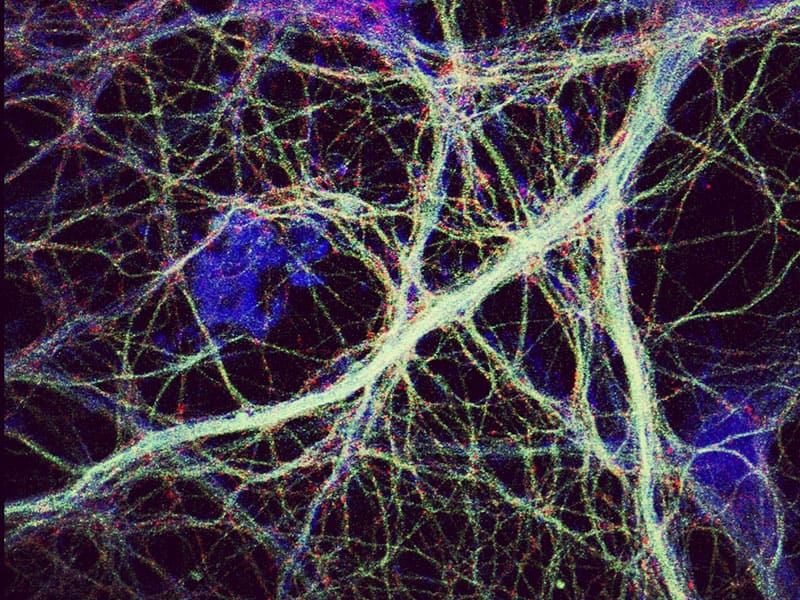
Pyramidal Neuron from an Embryonic Rat Hippocampal Tissue
Anti-MAP2 antibodies (blue). alpha-Tubulin (green). BK channel alpha subunit (red).
Courtesy of Garrett E. Seale. Treistman Lab. UPR-Institute of Neurobiology
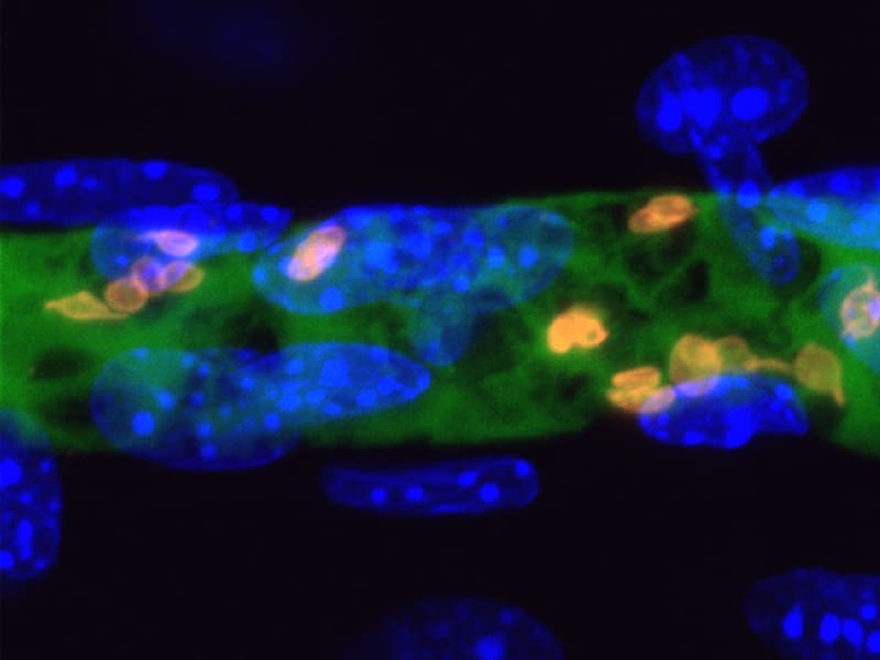
Vascular Endothelium from Mouse Cremaster Muscle Cells
Chimera composed of mouse TLT-1 and human Fc/IgG stained with FITC-conjugated anti-human IgG (green). TLT-1 stained with (primary antibody) rabbit anti-mouse TLT-1 and (secondary antibody) Dylight 649-conjugated donkey anti-rabbit antibody (orange). Nuclei stained with DAPI (blue).
Courtesy of Jessica Morales. Washington Lab. UPR-Río Piedras Biology
Mouse Cremaster Muscle Cells at Late Stages of Platelet Activation
Neutrophils stained with FITC-conjugated anti-GR-1 antibody (green). CD41 glycoprotein stained with (primary antibody) PE (phycoerythrin)-conjugated anti-CD41 antibody (red). Nuclei stained with DAPI (blue). TLT-1 stained with (primary antibody) rabbit anti-mouse TLT-1 and (secondary antibody) Dylight 649-conjugated donkey anti-rabbit antibody (yellow).
Courtesy of Jessica Morales. Washington Lab. UPR-Río Piedras Biology

Proximal End from Intestinal Mesentery of a Sea Cucumber
Nervous tissue stained with mouse antiserum RN1 antibody (red).
Courtesy of Christian Nieves. García Arraras Lab. UPR-Río Piedras Biology
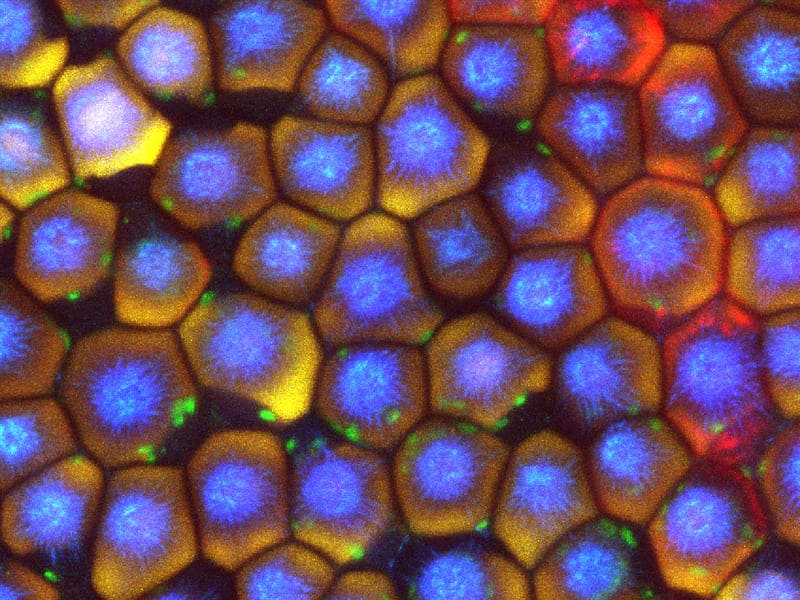
Plant Cells from an Orange Rose Petal
Auto-fluorescence of petal cells at maximum intensity projection (MIP). Image was taken with four different excitation lasers: 405 nm (blue), 488 nm (green), 561 nm (yellow), and 640 nm (red).
Courtesy of Bismark Madera-Soto. Lasalde Lab. UPR-Río Piedras Biology
Fasciola hepatica Adult Worm
FhTP16.5 protein labelled with (primary antibody) rabbit anti-FhTP16.5 IgG and (secondary antibody) FITC-conjugated anti-rabbit IgG (green), TRITC-conjugated phalloidin (red), Nucleus stained with DAPI dye (blue).
Courtesy of José F. Gaudier-Pagán. Espino Lab. UPR-Medical Sciences Microbiology
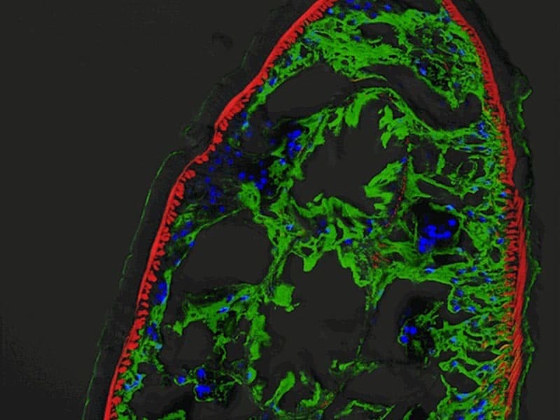
Hair Cells from Zebrafish Neuromasts
Membrane-targeted green gluorescence protein (mGFP, cyan-white gradient) driven by Brn3c promoter/enhancer. Differentiated hair cells (violet) in the sensory organ tissue from a Brn3C:mGFP transgenic zebrafish.
Courtesy of Luis Colón. Behra Lab. UPR-Medical Sciences Anatomy and Neurobiology
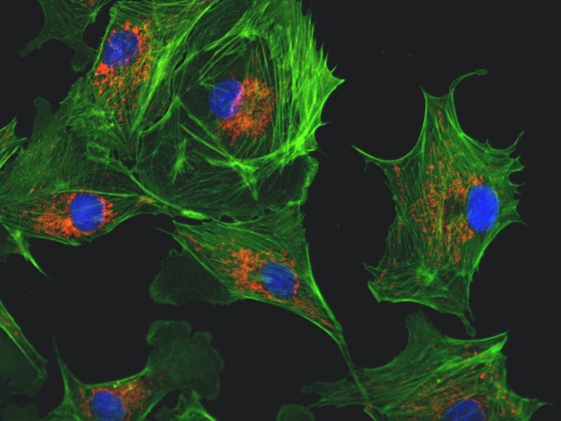
Bovine Pulmonary Artery Endothelial Cells
DNA stained with DAPI (4',6-diamidino-2-phenylindole) dye (blue). F-actin stained with phalloidin peptide conjugated to fluorescent Alexa Fluor 488 dye (green). Mitochondria stained with MitoTracker Red CMXRos dye (red).
Courtesy of Bismark Madera-Soto. Lasalde Lab. UPR-Río Piedras Biology

MRC-5, Human Fetal Lung Fibroblast Cells
Transferrin protein conjugated to FITC (fluorescein isothiocyanate) dye (green). Plasma membrane stained with phalloidin conjugated to TRITC (tetramethylrhodamine B isothiocyanate) dye (red). DNA stained with DAPI dye (blue).
Courtesy of Zally Torres. Griebenow Lab. UPR-Río Piedras Chemistry
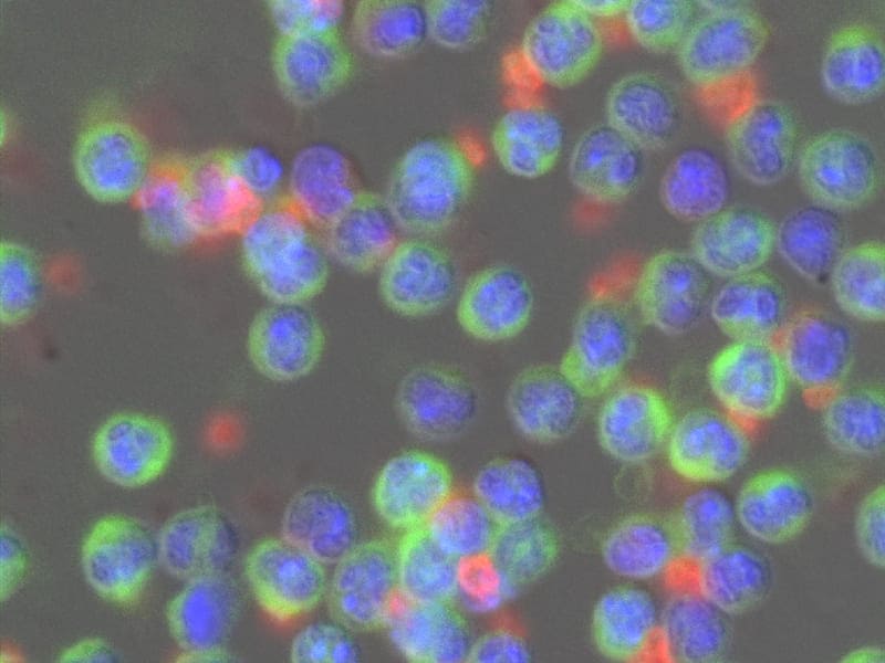
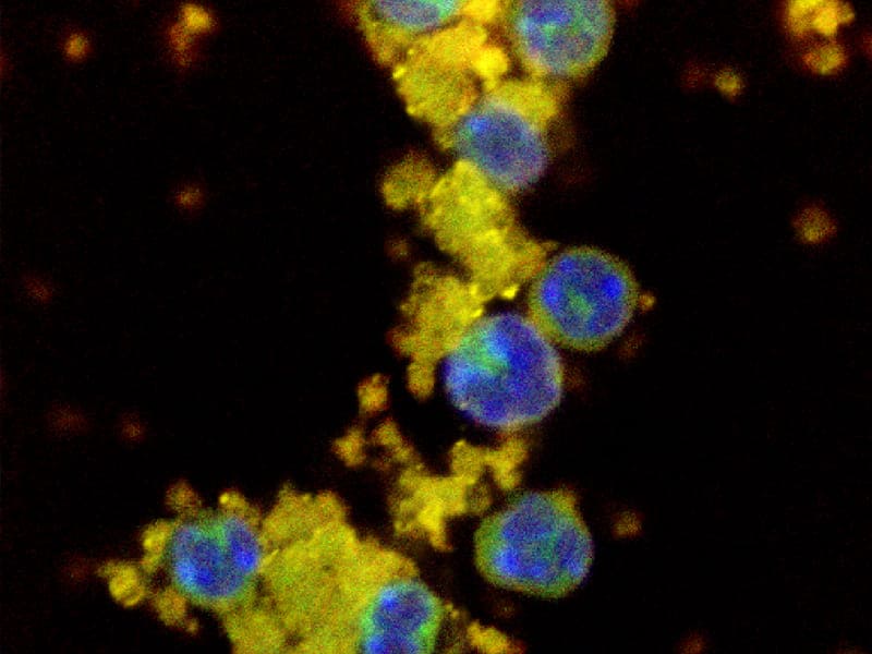
Mouse Bone Marrow Polymorphonuclear Cells and Thrombin Activated Platelets
Nuclei stained with DAPI (blue). Neutrophils stained with FITC-conjugated anti-GR-1 antibody (green). CD41 glycoprotein stained with (primary antibody) PE-conjugated anti-CD41 antibody (red).
Courtesy of Jessica Morales. Washington Lab. UPR-Río Piedras Biology
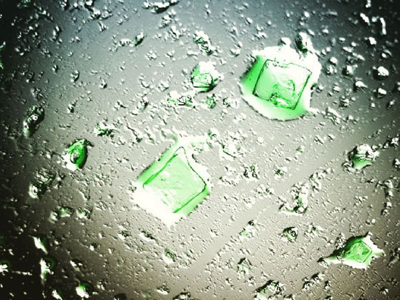
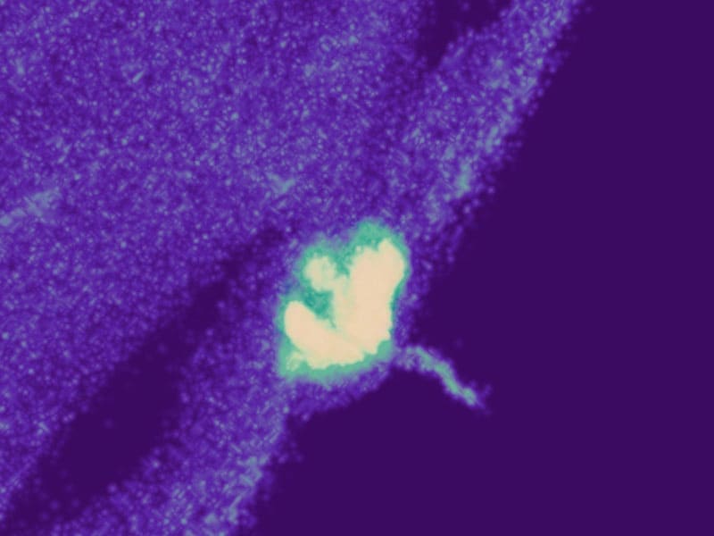
Nicotinic Acetylcholine Receptor Crystals Isolated from Torpedo Electroplax
Nicotinic Acetylcholine Receptor (nAChR) stained with α-bungatotoxin protein conjugated to fluorescent Alexa Fluor 488 dye (green). nAChR isolated from electric organ (electroplax) of the electric ray (Torpedo californica).
Courtesy of Luis F. Padilla. Lasalde Lab. UPR-Río Piedras Biology